
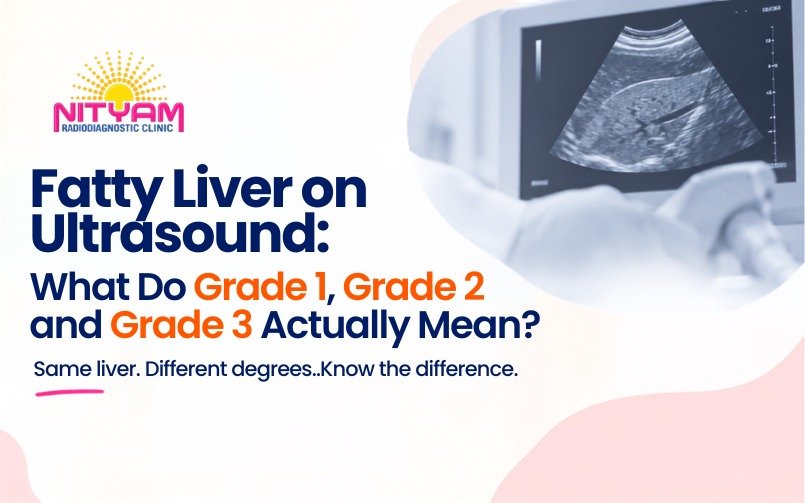
Quick Answer: Understanding the Three Grades
When an ultrasound report mentions Grade 1, Grade 2, or Grade 3 fatty liver, it describes the precise degree of fatty change visible in the liver on imaging. In simple terms, Grade 1 means mild fatty change, Grade 2 means moderate fatty change, and Grade 3 means severe fatty change.
However, patients frequently misunderstand the word “grade.” It does not mean Grade 1, Grade 2, and Grade 3 represent three stages of liver failure. A higher ultrasound grade mainly indicates that the liver appears increasingly bright on the monitor and that deeper liver structures have become harder for the radiologist to visualize.
Most importantly, an ultrasound fatty liver grade does not, by itself, tell you how much liver scarring or fibrosis is currently present. That distinction matters immensely because fat, inflammation, fibrosis, and cirrhosis are completely different medical conditions. Therefore, if your report says “fatty liver,” the right response is not to panic. Instead, you should understand what the finding means, consider the possible causes, and discuss whether additional evaluation suits your health profile.
Introduction: What Does “Fatty Liver” Actually Mean?
You may have gone for an abdominal ultrasound because of stomach discomfort, a routine health check, or another specific medical concern. Then, while reading the final report, you noticed an unexpected sentence: “Grade 1 fatty liver,” “Grade 2 fatty liver,” or “Grade 3 fatty liver.”
Naturally, one question immediately follows: “How serious is my liver condition?”
Fatty liver, medically called hepatic steatosis, occurs when excess fat actively accumulates inside the liver cells. It remains a highly common finding during abdominal imaging, and many people carry it without experiencing any obvious physical symptoms.
On a conventional ultrasound, fat fundamentally changes the way sound waves travel through the liver tissue. Consequently, the liver appears brighter than normal. As the amount of fat increases, deeper internal structures become far less clearly visible. Radiologists utilize these specific visual features to accurately describe the degree of steatosis. This means the grade on your ultrasound provides highly useful information, but it represents only one part of your clinical picture. You should never interpret it as a complete, final measure of your overall liver health.
Table of Contents
- What is Fatty Liver?
- What Do Grade 1, Grade 2 and Grade 3 Mean?
- How Does Ultrasound Detect Fatty Liver?
- What Else Can an Abdominal Ultrasound Evaluate?
- Does the Ultrasound Grade Tell You About Liver Damage?
- What Can Contribute to Fatty Liver?
- What Should You Do After Fatty Liver Is Found?
- Can Fatty Liver Improve?
- When Should You Speak to a Doctor?
- Frequently Asked Questions
What is Fatty Liver?
Fatty liver means that an abnormal, unhealthy amount of fat has accumulated directly within your liver cells.
Currently, doctors call the most common metabolic form MASLD, or metabolic dysfunction-associated steatotic liver disease. Physicians often associate this condition with metabolic problems such as excess body weight, abdominal obesity, diabetes, insulin resistance, high cholesterol, high triglycerides, high blood pressure, and low physical activity.
Additionally, alcohol consumption heavily causes fatty changes in the liver. Certain medicines and other rare medical conditions contribute as well. Therefore, the exact same ultrasound finding can stem from completely different root causes in different people. One vital point is that you might have fatty liver without showing any obvious symptoms. Many people feel completely well and discover the condition only because a doctor ordered an abdominal ultrasound for a totally unrelated reason. For this reason, an ultrasound report provides a brilliant, early opportunity to recognize a hidden metabolic concern.
What Do Grade 1, Grade 2 and Grade 3 Mean?
You can easily remember the conventional ultrasound grading system as follows:
- Grade 1 — Mild fatty liver
- Grade 2 — Moderate fatty liver
- Grade 3 — Severe fatty liver
Radiologists base these grades mainly on the liver’s brightness (increased echogenicity) and on how much the fatty change visually obscures vessels and deeper structures. Therefore, these grades represent a visual assessment of steatosis, not a precise measurement of liver fat percentage.
Grade 1 Fatty Liver: Mild
In Grade 1, the liver usually appears mildly brighter than expected on the monitor. Important structures and deeper areas generally remain fully visible. Doctors consider this mild hepatic steatosis, and it does not automatically mean permanent damage exists. However, you should not dismiss it. A mild finding strongly associates with metabolic risk factors, providing an excellent reason to review your weight, blood sugar, and diet.
Grade 2 Fatty Liver: Moderate
In Grade 2, the liver appears much more noticeably bright. The increasing echogenicity makes intrahepatic blood vessels less distinct and deeper portions significantly more difficult to visualize. While Grade 2 represents more pronounced steatosis than Grade 1, it still does not automatically mean you have cirrhosis or advanced scarring. Your doctor will consider this finding alongside your medical history and blood tests.
Grade 3 Fatty Liver: Severe
Grade 3 acts as the most marked category in conventional ultrasound grading. The liver appears remarkably bright, and stronger attenuation of the ultrasound beam drastically reduces the visualization of deeper structures. Consequently, blood vessels and the diaphragm become incredibly difficult to see clearly. This finding absolutely deserves appropriate medical attention. Yet, you must not misunderstand the terminology: Grade 3 means severe fatty change is visible, but it does not automatically equal cirrhosis.
How Does Ultrasound Detect Fatty Liver?
An ultrasound machine does not simply count how many grams of fat reside inside the liver. Instead, the radiologist expertly evaluates the liver’s visual appearance during the live examination.
As fat accumulates, the liver generally becomes more echogenic (it appears brighter on the image). With increasing steatosis, the fat increasingly attenuates the ultrasound beam as it travels through the tissue. In simple terms: More fatty change → brighter liver → greater beam attenuation → deeper structures become less clearly visible.
Conventional abdominal ultrasound remains highly practical, widely available, completely non-invasive, and completely free from ionizing radiation. However, ultrasound carries limitations. Mild amounts of liver fat occasionally prove hard to detect, and image quality varies with patient factors. Therefore, ultrasound findings are interpreted alongside clinical parameters and technical factors to ensure standardized radiological evaluation.
What Else Can an Abdominal Ultrasound Evaluate?
An abdominal ultrasound goes far beyond merely looking for fatty liver. During the same examination, the radiologist actively assesses several vital organs throughout your abdomen.
- The Gallbladder: Examined for hidden stones, wall thickening, or inflammation.
- The Bile Ducts: Evaluated for abnormal dilatation or blockages.
- The Pancreas: Assessed thoroughly when adequately visualized.
- The Spleen and Kidneys: Checked for size, structural changes, stones, and cysts.
Furthermore, the radiologist looks for free fluid within the abdomen. Because patients often possess more than one abdominal finding simultaneously, reading the report as a complete examination provides the most value.
Does the Ultrasound Grade Tell You About Liver Damage?
This is perhaps the single most important question patients ask. The straightforward answer is: Not by itself.
The ultrasound grade strictly describes the visual severity of steatosis. It does not directly measure cellular inflammation, fibrosis, or cirrhosis. A person can carry substantial steatosis without having advanced fibrosis. Conversely, another person might have important fibrosis not fully explained by the fatty change seen on screen. Consequently, doctors often use specialized blood tests and non-invasive fibrosis assessments (like FIB-4 and elastography) to gather additional, critical information.
What Can Contribute to Fatty Liver?
Fatty liver develops for several complex reasons, making identifying the underlying contributor crucial to your recovery.
- Excess Weight and Abdominal Fat: Being overweight heavily associates with metabolic dysfunction.
- Diabetes and Insulin Resistance: When your body struggles to regulate blood sugar, fat accumulation risks soar.
- High Cholesterol and Triglycerides: Abnormal blood lipid levels directly drive metabolic liver conditions.
- Dietary Habits: High-calorie diets, sugary drinks, and heavily processed foods overwhelm the liver’s processing capacity.
- Low Physical Activity: A sedentary lifestyle makes maintaining healthy blood sugar extremely difficult.
- Alcohol: Regular alcohol consumption causes direct fatty changes, which often overlap dangerously with metabolic disease.
What Should You Do After Fatty Liver Is Found?
If your report highlights a fatty liver, your immediate next step involves determining the specific root causes alongside your doctor. They will likely review:
- Liver Blood Tests: Checking liver enzymes (though normal enzymes do not completely exclude disease).
- Blood Sugar: Running fasting glucose or HbA1c tests to identify diabetes.
- Cholesterol and Triglycerides: Mapping your overall cardiovascular risk.
- Fibrosis Risk: Calculating a FIB-4 score to evaluate potential scarring.
Can Fatty Liver Improve?
Yes. In many people, liver fat actively improves when they address the contributing metabolic and lifestyle factors. Regular physical activity profoundly supports metabolic health. Similarly, healthy eating helps control excess calorie intake, improving blood sugar and lipid levels. For overweight patients, medically appropriate weight reduction dramatically reduces liver fat. The earlier you recognize the underlying risk factors, the easier you can take sensible, highly effective steps toward better long-term health.
When Should You Speak to a Doctor?
A fatty liver finding rarely dictates emergency treatment. However, you absolutely must discuss the result with your doctor—particularly if you have diabetes, obesity, high cholesterol, abnormal liver enzymes, or a history of regular alcohol consumption.
You should seek urgent medical attention only if you experience severe symptoms such as intense abdominal pain, vomiting of blood, black stools, severe confusion, or fainting. The ultimate purpose of evaluating fatty liver is never simply to “lower the ultrasound grade.” The true goal involves identifying the cause, understanding the long-term risk, and actively protecting your liver.
Frequently Asked Questions
- 1. Is Grade 1 fatty liver dangerous?
- Grade 1 usually indicates mild steatosis and does not automatically mean serious liver damage. However, it is still worth reviewing possible metabolic causes and discussing the finding with your healthcare professional.
- 2. Is Grade 2 fatty liver serious?
- Grade 2 means moderate fatty change on ultrasound. It does not automatically indicate cirrhosis, but you should consider the finding alongside other health and metabolic risk factors.
- 3. Does Grade 3 fatty liver mean cirrhosis?
- No. Grade 3 indicates severe fatty change visible on a conventional ultrasound. It does not, by itself, determine the exact stage of liver fibrosis or prove that cirrhosis is actively present.
- 4. Can a person with normal liver tests still have fatty liver?
- Yes. Liver enzymes can sometimes remain completely normal even when fatty liver is heavily present. Imaging and blood tests provide entirely different types of medical information.
- 5. Can thin people get fatty liver?
- Yes. Fatty liver is not strictly limited to people who are overweight. Metabolic factors, genetics, alcohol, medicines, and other medical conditions play massive roles regardless of body size.
- 6. Can Grade 1 fatty liver be reversed?
- Liver fat can improve dramatically in many people when they appropriately address the underlying causes and metabolic risk factors. The right approach always depends on the individual.
- 7. Is ultrasound enough to measure liver damage?
- No, not always. Conventional ultrasound provides highly useful data for identifying and grading fatty change, but it does not provide a complete, microscopic assessment of liver fibrosis or cellular inflammation.
- 8. Do I need a FibroScan if fatty liver is found?
- Not everyone needs elastography. Your doctor will recommend it depending strictly on your blood tests, medical history, risk factors, and overall clinical assessment.
- 9. Can fatty liver cause symptoms?
- Many people experience absolutely no obvious symptoms. When symptoms do occur, they usually remain quite non-specific, which is exactly why doctors frequently discover fatty liver incidentally during routine imaging.
- 10. Should I be worried if my report says Grade 3?
- You should take the finding seriously, but you should not assume it means your liver has failed. Grade 3 describes the ultrasound appearance of severe steatosis. Your doctor can determine whether additional testing is required.
Diagnostic Services Information
Nityam Radiodiagnostic Clinic provides clinical diagnostic imaging services including CT Scan, Ultrasound & Sonography, Color Doppler, Digital X-Ray, Portable X-Ray, Mammography, OPG Dental X-Ray, and Image-Guided Interventionss. The clinic performs these services under standardized clinical protocols upon medical referral. In accordance with ethical guidelines, clinical establishments may communicate objective and verifiable particulars concerning their establishment. This includes contact details, diagnostic facilities, and the availability of doctors.
In compliance with National Medical Commission guidelines, clinical establishments and Registered Medical Practitioners publishing electronic media posts must transparently disclose the practitioner’s name, qualifications, registration status, and State Medical Register (SMR) or National Medical Register (NMR) number. This educational material serves public awareness. It does not promote personal practice, solicit patients, or make unverifiable medical claims.
By Dr. Chirag Arvindkumar Nayak
Qualifications: MBBS, MD, DMRE, PDCC
Registration Status: Consultant Radiologist | Founder & Head
SMR Registration Numbers: G-30018 (MBBS) | G-13167 (MD) | G-12404 (DMRE)
Nityam Radiodiagnostic Clinic
4 to 8, Ground Floor, Samved Complex,
Jail Road, Devbagh, Bhavnagar, Gujarat – 364001
Phone: +91 9427060235
Email: info@nityamradiologyclinic.com
Website: nityamradiologyclinic.com
This article is intended for general educational and informational purposes only. It does not replace personalized medical advice, clinical examination, diagnosis, or treatment from a qualified healthcare professional.